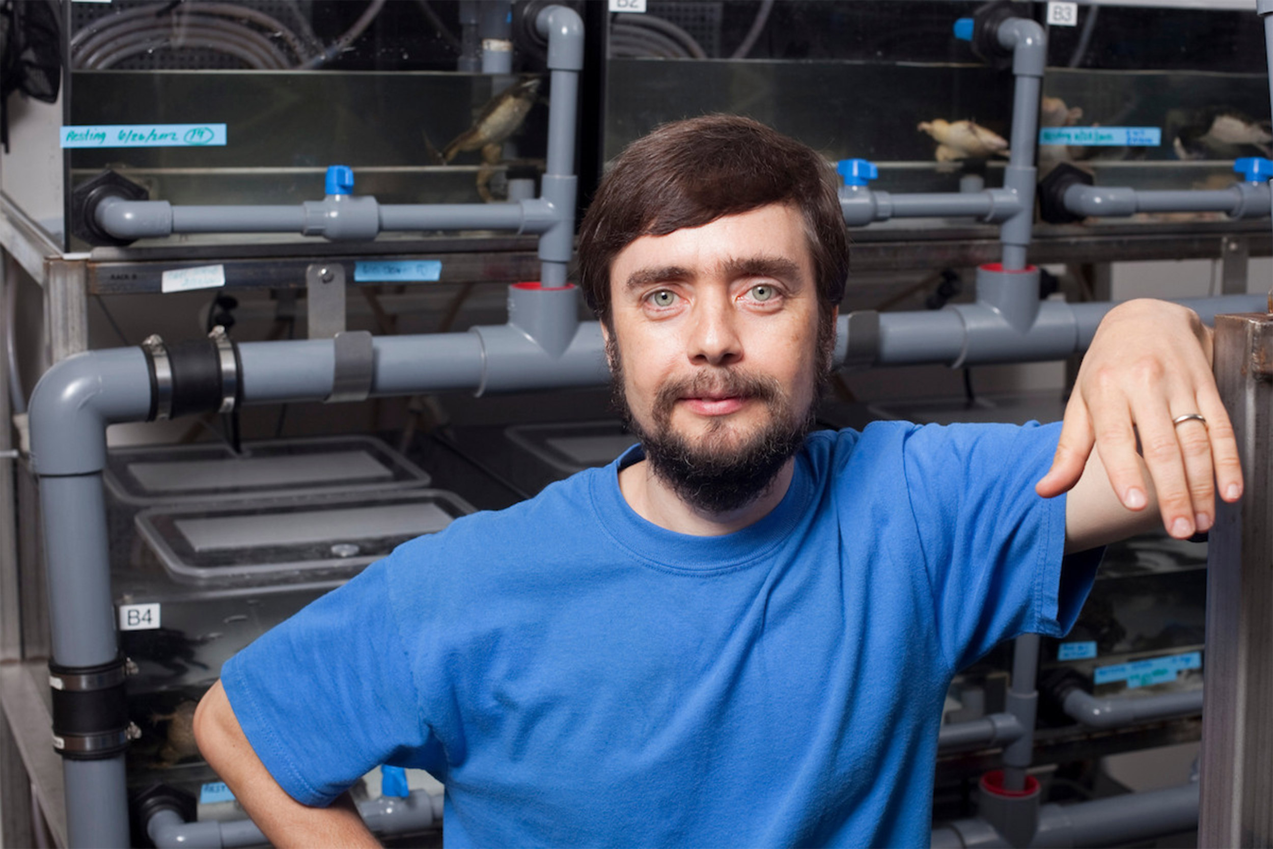
“I believe the capacity to create and repair specific structures is evolutionarily ancient and highly conserved across the tree of life, and that there is no reason it can’t someday be activated in human patients,” said Mike Levin, associate faculty member at the Wyss Institute.
Credit: Wyss Institute at Harvard University
Electrifying insights into how bodies form
Mike Levin studies how cells make collective decisions about growth and shape using bioelectricity
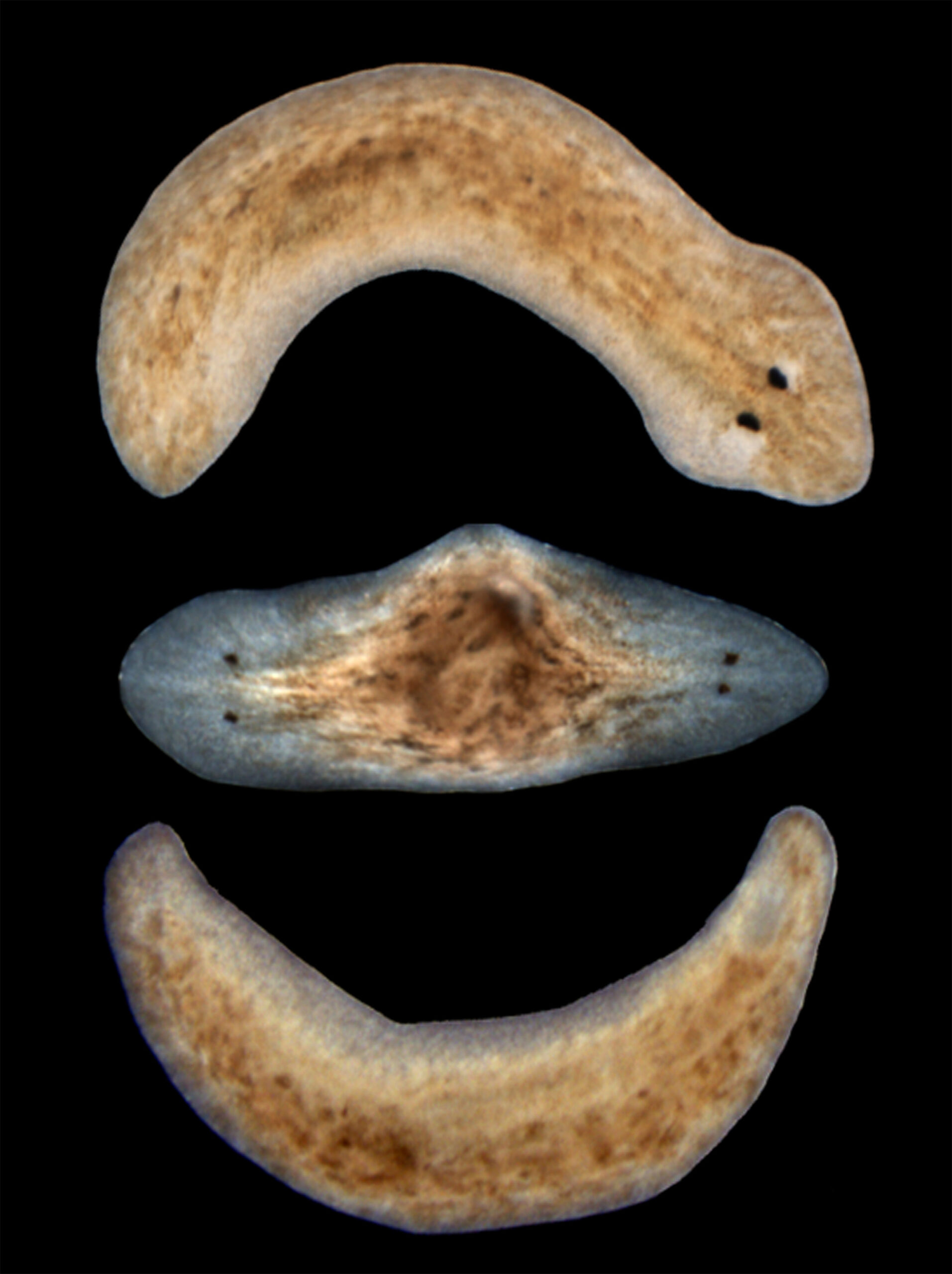
At first glance, Mike Levin’s lab looks like any standard biology lab with its shelves of petri dishes containing small, brown, semitransparent flatworms called planaria, one of the organisms his lab studies. Look more closely at the bodies of the tiny worms swimming and stretching under the plastic, however, and you might notice something strange — instead of a head and tail, each worm has two fully functional heads.
Planaria are the champions of regeneration: Chop one into multiple pieces, and each fragment will regrow exactly the parts it needs to transform into a perfect tiny worm, no more, no less. How do the cells in each worm fragment know what’s missing, and how to rebuild the organs it needs, and when to stop growing? Levin’s group at Harvard’s Wyss Institute for Biologically Inspired Engineering is trying to answer these questions, because he believes that doing so is the key to future advances in regenerative medicine and synthetic bioengineering. While biology has started to identify the genes and proteins involved in regeneration, how and when cells use those tools to build complex anatomical features remains unknown.
Levin and his team are tackling this challenge by trying to figure out how cells within a worm fragment coordinate to create a specific structure, and then manipulating those cellular conversations to change how the fragments regenerate — with a head at each end, for example. Remarkably, their research has shown that just one brief, conversation-changing treatment is enough to make regenerating planaria continue to produce two-headed worms in subsequent rounds of regeneration, without any further manipulations. Surprisingly, this permanent change in anatomy is produced not by editing the worms’ genes but by targeting a different aspect of biology that is attracting renewed attention after being long ignored: bioelectricity.

As far back as the 18th century, scientists realized that applying electrical currents to dead animals could make their muscles twitch, and the idea that electricity was the literal “spark of life” caught on. It was such a pervasive theory that it made its way into literature and the arts, exemplified by Mary Shelley’s 1818 novel “Frankenstein,” in which a lightning storm inspires the young Dr. Frankenstein to bring the dead to life. But the actual scientific study of bioelectric signals proved to be extremely challenging, because the moment an organism is plucked, dried, fixed, or preserved, the electricity vanishes.
“When cells and tissues are alive, there’s a bioelectric potential between the inside of a cell and the outside,” Levin said in a recent interview in his lab. “As soon as that potential collapses, the cell is dead. So, I think it’s fair to say that bioelectricity is the spark of life. But more importantly, the bioelectric potential is not just a byproduct of living; it is a medium that cells exploit to communicate with each other and to form networks that are much more than the sum of their parts.”
Because bioelectrical studies can be performed only on living cells, other analytical methods that work in fixed tissues and fractionated cells surged ahead in popularity during the 20th century, as techniques like biochemistry and molecular genetics began to reveal the complex choreography of molecules that direct cellular behavior. Electricity remained a central focus in niche fields such as neuroscience and cardiology, as the transmission of electric impulses was known to be the signaling method of choice along neurons and in the heart’s “pacemaker” nodes, but the idea that cells outside the nervous system communicate and are regulated by electrical signals fell out of fashion.
The body electric
When Levin was growing up in the 1980s, the boom in digital technology was ushering in a resurgence of interest in electricity in the form of personal computers. Levin was fascinated by the physics of electricity, and how it could be so readily harnessed to build circuits that perform computational functions. “The more I studied the use of electric circuits to implement memory and perform computation aimed at creating artificial intelligence, the more I thought that surely evolution must have found a way to exploit electricity for its capabilities long before brains showed up; cells and tissues had to start making a lot of complex decisions all the way back at the beginning of multicellular life,” he said.
The thought remained just an idea until a day in 1986 when Levin attended the Expo 86 World’s Fair in Vancouver, British Columbia. But the moment that came to define his career didn’t happen in one of the pavilions showcasing the world’s newest technologies — it happened in a used book shop, where Levin happened to pick up the “The Body Electric,” published the previous year by orthopedic surgeon Robert O. Becker.
“What was remarkable about that book was that it cited all these older research papers in which people had actually found evidence of electrical signaling outside of the nervous system during embryogenesis and regeneration, not only in animals but also in plants and fungi and other organisms,” Levin recalled. “I had never seen it in any modern textbook, but the fact that these studies were out there suggested that evolution really did discover how good the biophysics of electricity is for computing and processing information in non-neural tissues, and that might be a very interesting direction for understanding how cells cooperate to make decisions about constructing and repairing complex body parts.”
Levin has been moving in that direction ever since, earning dual B.S. degrees in computer science and biology at Tufts University and a Ph.D. from Harvard’s Graduate School of Arts and Sciences, followed by postdoctoral work at Harvard Medical School, where he began to uncover a new bioelectric language by which cells coordinate their activity during embryogenesis. He has continued and expanded that work in his own lab, first at Harvard’s Forsyth Institute, and now at the Allen Discovery Center at Tufts University and the Wyss Institute.
“My lab is looking at not only the mechanisms by which cells receive and transmit electrical signals, but how groups of all kinds of cells form distributed electrical networks that implement information processing,” Levin explained. “We’re interested in how tissues and organs compute using electrical signals — storing pattern memories and regulating large-scale anatomical remodeling. You can think about these groups of cells doing all the same things as a neural network, but everything goes at a much slower pace and is aimed at controlling cell behavior and anatomy, not muscles and body movement.”
Cracking the bioelectric code
Rather than the rapid-fire signals that travel through neurons to convey information like “this stove is hot” or “step on the brake,” the types of cellular bioelectric networks Levin studies direct much more complex processes, like “create an eyeball here,” “this side is your left,” or “heal this wound.” Over the last few decades, neuroscientists have developed techniques for recording the electrical signals that propagate through individual cells, which is very useful for studying neurons in the brain, but not sufficient for analyzing electricity on the scale of tissues or organs. In the last 15 years, Levin’s lab has been studying bioelectricity in a veritable zoo of animal models, including bacteria, slime molds, algae, frogs, human cultured cells, and of course, the planaria, to address that need.
“Understanding how tissues and organs encode and propagate anatomical information in electrical signals to fix or create very complex, specific structures is a fundamental challenge that we call ‘cracking the bioelectric code,’” said Levin. “It has the potential to advance not only biomedicine but multiple fields including robotics and AI, akin to how insights from neuroscience are being used to drive the development of neural nets and other computational tools, but in a much more general context.”
The discipline that could see the biggest benefit from cracking that code is the still-fledgling field of regenerative medicine — if scientists can figure out what series of electrical signals carries the instructions “build a leg here,” it could one day be possible to direct a human body to regrow a limb just like the planaria can regrow heads. “The ability to build a specific anatomical structure from different starting conditions, and stop when precisely the right pattern is finished, is one of the big, unsolved problems of developmental biology and regenerative medicine today,” said Levin. “Salamanders can regrow eyes, jaws, limbs, ovaries, and portions of their heart and brain. Deer — a large, adult mammal — regenerate antlers made of bone, nerve, and skin every year at a rate of about 1 centimeter of new bone per day. I believe the capacity to create and repair specific structures is evolutionarily ancient and highly conserved across the tree of life, and that there is no reason it can’t someday be activated in human patients.”

True to his background in computer science, Levin is incorporating machine learning and artificial intelligence into his lab’s efforts to uncover meaning in the vast amounts of functional data his research generates about anatomy and shape. His team builds AI-based tools that can mine those data to build models that help them understand how cells and tissues make decisions to repair and create specific structures, and to discover interventions that can manipulate that complex process. Recently, they created an AI system that analyzed hundreds of papers about planaria, digested information about the experiments that were performed on the worms, and discovered a novel way of thinking about the circuits that allow planaria to repair themselves, resulting in a new model of planarian regeneration — the first in this field that was not discovered by a human scientist.
Reorganizing the “lightning” in our cells
Now that innovations in recording and writing bioelectric information into living cells are making it feasible to study how electrical signals function in living organisms, there is renewed interest across many disciplines in how electricity interacts with other biological phenomena, such as genetics, metabolism, mechanobiology, and immunology. Since joining the faculty of Tufts University in 2009 as the Vannevar Bush Professor, Levin’s expertise has been sought for a number of multidisciplinary projects. In 2016, he was tapped to become the director of the Allen Discovery Center, recruiting 12 other investigators around the world who are combining their disparate backgrounds to understand and ultimately rewrite the “morphogenetic code” by determining where bioelectric patterns originate, how they map the organization of cells, and how their code is interpreted by cells’ genetic and molecular machinery to build and maintain an organism’s anatomy.
“We think that if we’re able to figure out these high-level controls of body plan and shape, we will be able to induce limb regeneration, birth defect repair, and tumor reprogramming, first in frogs and similar model systems, and later in mammals,” said Levin. “I would be incredibly excited if in the next decade we can move some of our discoveries toward the clinic, where they can actually help people.”
While Levin has collaborated with faculty at the Wyss Institute for more than a decade, his path to official membership also began in 2016, when the Defense Advanced Research Projects Agency (DARPA) funded called Technologies for Host Resilience (THoR), a multicenter project led by Wyss Institute Director Donald Ingber to investigate why some organisms tolerate pathogenic infection, and to uncover which biological mechanisms are responsible for their resilience. The Wyss Institute was chosen to lead the interdisciplinary project, and Levin was invited to join as an associate faculty member in 2017. His role in this project was to investigate bioelectricity’s impact on immunity, collaborating with Ingber, core faculty member James Collins, and lead senior staff scientist Michael Super.
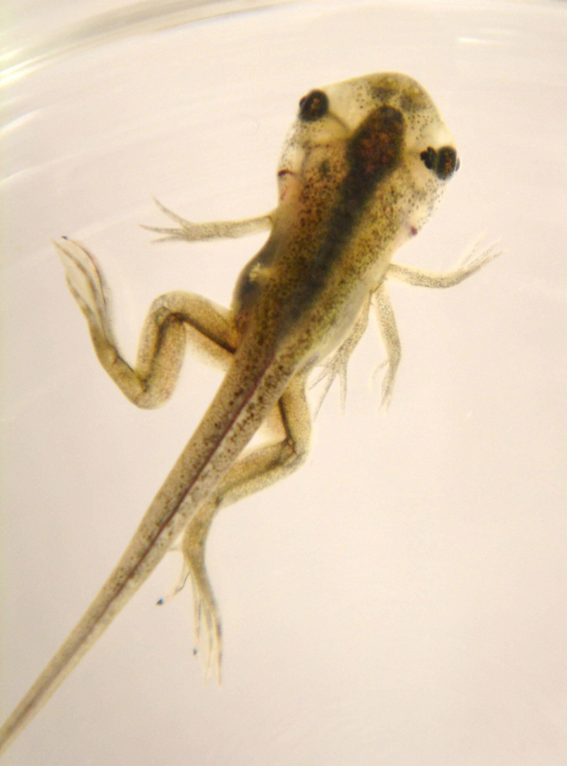
Early results of his research revealed that administering drugs that target ion channel proteins to make cells’ interiors more negatively charged strengthened tadpoles’ innate immune response to E. coli infection and injury, suggesting that the immune system is regulated by non-neural bioelectricity. The change in voltage altered the expression of genes in the tadpoles that are also involved in human immune responses, indicating that modulating bioelectric charge could be a new clinical approach to reducing infections.
Most recently, Levin has started working on the Wyss’ Biostasis Project, a similarly large, cross-functional and interdisciplinary effort also led by Ingber whose goal sounds like something out of science fiction: find a way to slow down biological time. “A lot of my lab’s prior work has focused on looking at how organisms arrange themselves in 3D space, but time is equally important to living creatures,” Levin said. “With this new project, we’re shifting to ask how the different physiological, genetic, and anatomical structures in an animal keep time, and how we might alter the rates of these various processes for improved survival, repair, and health.”

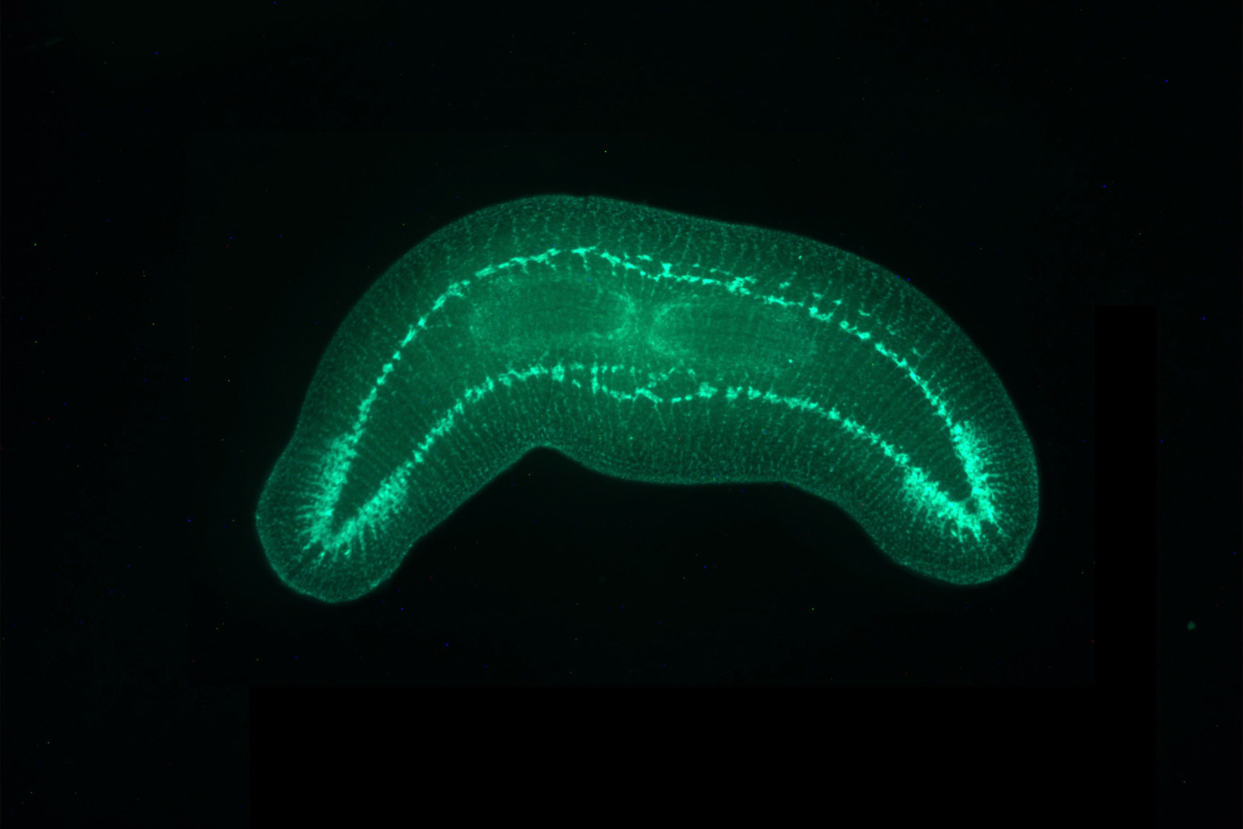
Tools are being developed for visualizing the interplay of bioelectrical signals between cells, such as this technique that labels synapsins (proteins that are present in the synapses of neurons) in a double-headed worm.
Credit: Mike Levin/Tufts University
To answer those questions, his lab is developing new constructs that can report biological time, such as fluorescent molecules that indicate metabolic, proliferative, bioelectric, and circadian cycles during development and regenerative healing. The researchers are also testing candidate compounds that could potentially halt or slow biological time, and in the future they will be mining some of the novel model systems they have developed for new compounds that cells and organisms use to regulate their own and each other’s experience of time.
Levin’s research has come a long way from his initial fascination with electrical circuits in computers, and the diversity of projects he’s involved in reflects how bioelectricity is reclaiming its place alongside genes, proteins, and mechanical forces as an important piece of the puzzle that is figuring out how life works.
“I think all the different projects that my lab is working on link together in an effort to answer the question, ‘How do biological systems store and process spatial and temporal information to behave adaptively, build large functional structures, and resist challenge?’” said Levin.


