New study pairs immunolabeling with connectomic data

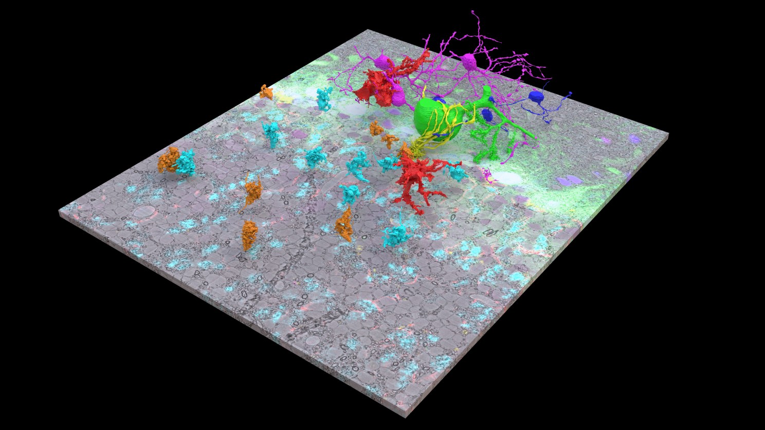
An electron microscopy image volume from a cerebellar cortex overlaid with six colors of protein labeling by scFv probes. The floating colorful objects are cells and cellular structures.
Credit: Xiaomeng Han
Harvard researchers have developed a technique for adding color-coded localization data for pinpointing different biomolecules to high-resolution images of a mouse brain.
Publishing in Nature Communications, first author Xiaomeng Han and colleagues used glowing molecular probes that bind to specific proteins to create a colorful overlay for connectomic electron microscopy data. The combined data allowed them to observe new details in a sample from a mouse cerebellar cortex.
“Neural circuits can be mapped using serial section electron microscopy technique, but these do not provide information about cell types or other molecular annotations,” said senior author Jeff Lichtman, the Jeremy R. Knowles Professor of Molecular and Cellular Biology and dean of science in the Faculty of Arts and Sciences. “This work describes an approach to superimpose molecular annotations on serial electron microscopy volumes by taking advantage of the high permeability of small fluorescent antibody fragments.”
In the study, the scientists used a type of immunolabeling probe called single chain variable fragments. They are derived from monoclonal antibodies but are much smaller and can penetrate deeper into tissue.
The researchers collected data from a mouse cerebellum labeled with five different scFv probes, each of which detected a different protein. Working with Doug Richardson and the Harvard Center for Biological Imaging, the researchers used spectral unmixing to capture six (five from scFv labeling, one from cell nuclei labeling) distinct fluorescent signatures. Using that fluorescence data, Han and colleagues created a colorful fluorescence overlay for the electron microscopy data.
Analysis of the combined dataset revealed several unexpected details about the mouse cerebellar cortex. For example, the researchers were able to identify a rarely-studied type of cell called a molecular layer granular cell. They were also able to localize a type of voltage-gated potassium channel at cellular subcompartments called juxtaparanodes. Finally, they were able to distinguish two molecularly defined, mossy fiber terminals that use different neurotransmitter transporters.
Han and colleagues have made their scFv probes available to other researchers through Addgene in hopes that the technique will be replicated in other connectomics studies.
The study was a collaboration of the Lichtman lab and James Trimmer’s lab at University of California Davis.


